Wednesday, April 21, 2010

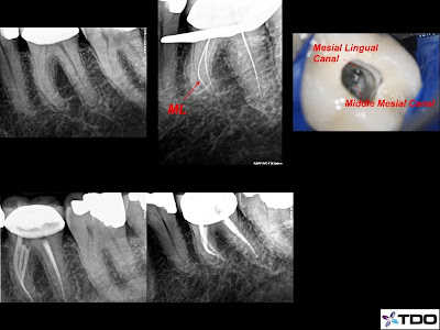
Retreatment with missed canals


This patient presented with severe pain.
Root canal retreatment was initiated on tooth number 30. Her symptoms disappeared after the first visit, after locating and instrumenting the missed DL canal. There was also a Middle Mesial canal.
Interestingly, the final radiograph doesn't look that different.

Tuesday, April 6, 2010

Best Of Baltimore 2010

Every year Baltimore Magazine surveys 2000 dentists in Baltimore and the surrounding areas for their feature article 'Baltimore's Top Dentists'.

We are proud and honored to be listed as a top endodontist in Howard County. Heartfelt thanks go out to all of our peers who recognized the hard work that we do everyday saving teeth and alleviating pain for their patients.

It is a wonderful feeling to be recognized by the amazing dentists who practice in this area. We will continue to exceed the expectations of our referring doctors and their patients, for many more years.

Wednesday, April 15, 2009

Sedation Dentistry at its Best

This is a case I am particularly proud of.

This patient came into my office with an extreme case of dental phobia and claustrophobia. The very thought of wearing a rubber dam terrified her. With a gentle explanation of the procedure and administration of oral sedation I completed a successful and anxiety free root canal treatment.

Doctors, check out the THREE joining Mesial Canals!


Monday, February 16, 2009

Invasive Cervical Resorption

Invasive Cervical Resorption is a relatively uncommon and often misdiagnosed form of external root resorption. This type of resorption is generally insidious and is often misdiagnosed as a type of internal root resorption. Unfortunately this misdiagnosis often leads to the loss of the tooth.

Invasive cervical resorption has four classifications:

Class I is characterized as having a small lesion near the cervical area with shallow penetration into the dentin. You may observe a defect containing tissue that bleeds upon probing.
Class II has a well defined lesion that has penetrated close to the coronal pulp. You may begin to see a pink color in the crown of the tooth due to vascular tissue that is visible through thinning enamel. Upon radiological examination you will see a radiopaque line surrounding the pulp space.
Class III denotes the extension of the lesion into the coronal 1/3 of the root. The lesion will have a 'moth eaten' appearance on a radiograph. Most cases are diagnosed during this stage.
Class IV denotes a large area of resorption beyond the coronal 1/3 of the root. Most class IV cases have an unfavorable prognosis.

Early detection is critical to the restorability of the tooth due to the aggressive nature of this condition. Dr. Jarboe has expertise in the diagnosis of invasive cervical resorption and has successfully treated several teeth.

Tuesday, January 6, 2009

Newest achievements to help our dental phobic patients

As you know we are totally committed to the quality of care and the comfort of our patients. Our commitment sometimes takes us to great lengths...literally.

Most recently, it took Dr. Jarboe to Atlanta, Georgia where she attended advanced IV sedation training from November 4th - 14th.

Taking ten days away from the office and even her family (including those adorable grandbabies) is the kind of thing that Dr. Jarboe is pleased to do when it provides a real benefit to the patients that we care so much about.

IV sedation allows patients who are particularly apprehensive to be most contented, as they are essentially able to relax through their procedures. Dr. Jarboe is the premier endodontist in the area to have completed this advanced training and we are in the process of leveraging this new option to make our patients even more comfortable.

Wednesday, October 22, 2008

Mac's Team


On Sunday, October 19 Dr. Jarboe and Dr. Ghahraman were pleased to attend the Bull Roast Fundraiser and to join Mac's Team in the fight against MLD. Mac is a four year old boy and the son of a very special patient of ours, who has been diagnosed with Metachromatic Leukodystrophy (MLD). MLD is a genetic condition that causes degeneration of the nervous system. Currently there is no cure for MLD, and the only treatments that are effective in slowing the course of this disease are bone marrow transplant and stem cell transplant. This little boy has shown more courage than most of us could ever find, and we are honored to have the opportunity to help in supporting Mac and his family. To learn more about MLD or to help in the efforts to facilitate improved research and treatments for this condition please visit Mac's web page at www.macsteam.org

Thursday, September 4, 2008

The use of digital photography is one of the latest innovations being implemented in our office. We have been using digital imagery for quite some time now, for several purposes. Taking a digital image of a tooth is a vital tool that we use to educate patients in their personalized treatment. We are able to document problems with teeth including, but not limited to, fractures, leakage, fistulas, and drainage of severely infected teeth. The greatest thing is that we are able to save these files and access them whenever we need to, with the simple click of a button. We find that our patients are particularly excited about the outcome, and are able to fully understand the complexity of treatment, when visual aides are introduced. We are proud to have the ability to showcase our work in the reports that we send out to the referring dentists, and the dentists appreciate having this detailed map of the procedure.

Because of the value that digital photography brings to our office, we aim to take the most precise images possible. Dr. Jarboe and Alicia recently attended a course in Evanston, Illinois in which they brought back valuable information about equipment and the latest photography techniques. We have installed the new equipment and immediately implemented the knowledge that they acquired in the course. In keeping with the standards of our office, we are continuing to send out more and more exemplary images with our reports.

Thursday, April 24, 2008

Rubber Dam

One of the most essential steps in successful endodontic treatment is the placement of the rubber dam.

It is the standard of care when performing root canal therapy. Without this key step it is impossible to maintain a sterile area, thus leading to potential contamination and ultimately the failure of the root canal treatment.

Not only does the rubber dam protect the tooth from bacterial contamination, it also serves as a protective barrier for the patient. The dam isolates the tooth being treated, helps prevent the leakage of irrigants into the oral cavity and can protect the patient from instruments such as hand files from falling into the oral cavity.

As you can see, our patient below is comfortable and happy to be wearing the rubber dam!(HA HA)

Wednesday, April 2, 2008

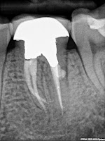
Perforation Repair

A 21 year old patient presented with a sinus tract associated with a previous root canal therapy on tooth number 19. The post perforated the distal root causing the failed treatment.


The root canal was retreated and the perforation site was repaired with MTA.


Check out the healing at 6 months!


Roya Ghahraman DDS